Mississippi INBRE Research Efforts Aided by Technology Upgrade
Fri, 06/18/2021 - 04:14pm | By: David Tisdale
 An enhancement of the cutting edge technology employed by the Mississippi INBRE’s
(IDeA Network of Biomedical Research Excellence) Imaging Facility, headquartered at
The University of Southern Mississippi (USM), will keep its affiliate faculty and
student researchers at the forefront in STEM (Science, Technology Engineering and
Mathematics) research.
An enhancement of the cutting edge technology employed by the Mississippi INBRE’s
(IDeA Network of Biomedical Research Excellence) Imaging Facility, headquartered at
The University of Southern Mississippi (USM), will keep its affiliate faculty and
student researchers at the forefront in STEM (Science, Technology Engineering and
Mathematics) research.
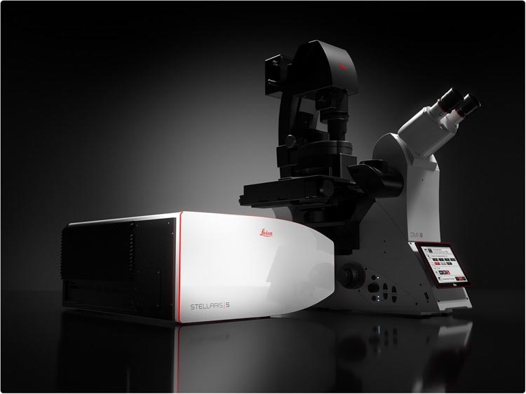
The Mississippi INBRE Imaging Core has upgraded its existing Leica SP8 confocal microscope to the STELLARIS STED super resolution platform, one of the most complete imaging systems in the region. The instrument was acquired through funding from the NIH-supported Mississippi INBRE Imaging Core Facility, as well as an NSF Major Research Instrumentation Program grant whose Principal Investigator (PI) is Dr. Alex Flynt, an associate professor in USM’s Center for Molecular and Cellular Biosciences.
The Mississippi INBRE Imaging Facility, directed by Dr. Jonathan Lindner, provides imaging and microscopy expertise to researchers throughout the state, offering access to and training on biomedical research equipment at no cost to users. The facility houses several types of microscopes, as well as a variety of large-scale instruments. The imaging facility also offers computational services and expertise to Mississippi researchers.
The addition of the STELLARIS STED super resolution microscope will enhance the quality and scope of biomedical research in the state of Mississippi, accommodating the varied needs of the INBRE Imaging Core user base. This cutting-edge technology is now accessible to faculty and students at USM and across the state who otherwise would not have access to super-resolution confocal microscopy.
 According to Dr. Flynt, while light-based microscopes are indispensable to the advancement
of many scientific fields. Unfortunately, he says, there is a lower limit to the size
of objects that can be observed due to the physics of light itself, a barrier that
impedes investigation of minuscule objects. Fortunately, recent advances have vaulted
over this hurdle, yielding “super-resolution” microscopes such as the STELLARIS STED.
According to Dr. Flynt, while light-based microscopes are indispensable to the advancement
of many scientific fields. Unfortunately, he says, there is a lower limit to the size
of objects that can be observed due to the physics of light itself, a barrier that
impedes investigation of minuscule objects. Fortunately, recent advances have vaulted
over this hurdle, yielding “super-resolution” microscopes such as the STELLARIS STED.
“This specific super-resolution technology is well-suited for imaging dynamic objects like those in cells, as well as nanoparticles created in the laboratory,” Dr. Flynt said. “Areas of research that will be investigated with this microscope include material scientists studying assembly of plastic-like materials, cell biologists, and biochemists investigating cell components important in Alzheimer’s and genetic tools, and microbiologists who examine bacterial community structures involved in infection and plant-soil interactions.”
Dr. Lindner concurs, noting also that researchers from a broad base of biological, chemical, and material science fields, including cellular and developmental biology, virology, biochemistry, high performance materials, and nanoparticle development, can greatly benefit from the instrument’s unique and powerful capabilities.
“Microscopes are essential tools for the investigation of biological and molecular systems,” Dr. Lindner said. “Access to cutting-edge instruments is vital for cell biology, embryology, biochemistry, and imaging advanced materials.
Further, the addition of the advanced microscope will provide important training opportunities for students, also enhancing Mississippi STEM education.
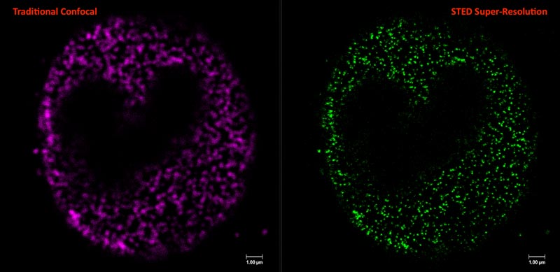
The Leica STELLARIS STED Super-Resolution Confocal Microscope upgrades the previous confocal microscope to a fully automated platform with a 3D STimulated Emission Depletion (STED) super resolution module. The STED technology enables fluorescence microscopy approaches for visualizing objects smaller than the diffraction limit of light, increasing resolution up to 10 times more than traditional microscopes. For reference, the diameter of a nucleus of an average human cell is approximately 10 micrometers. STED super-resolution imaging is capable of resolution below 50 nanometers, over 200 times smaller than a nucleus. This enables the real-time study of sub-cellular molecular interactions and mechanisms on the nanoscale.
The system is capable of both conventional confocal scanning and resonant scanning for rapid low-light illumination imaging, which is ideal for live specimens. It is equipped with an automated motorized stage with upgraded software for expanded view image stitching options, 3D modeling, FRAP, and co-localization. Additionally, the instrument is outfitted with an Okolab CO2 chamber for long term mammalian tissue culture imaging, and a Hamamatsu Flash camera for ultrafast acquisition.
Mississippi INBRE, directed by USM Professor Dr. Mohamed Elasri, is a statewide program supported by an award from the National Institutes of General Medical Sciences. Its mission is to enhance the biomedical foundation in Mississippi and engage talented researchers and students in biomedical research projects that will increase the state's research competitiveness, as well as positively impact the health of the state’s citizens.
For more information about Mississippi INBRE, visit msinbre.org.